REVO HR

Ultra-High Resolution. 3 µm.
See More. Diagnose Earlier.

REVO HR is an advanced all-in-one OCT system that combines ultra-high 3 µm resolution with a scanning speed of 130,000 A-scans per second. It provides comprehensive anterior and posterior segment diagnostics by integrating retinal OCT, AS-OCT, OCT-A, and a full-color fundus camera into a single platform.

Featuring UHR and UWF imaging technologies, it enables precise visualization of microstructures, detection of subtle pathological changes, and retinal assessment far beyond the macula. REVO HR sets a new standard in OCT imaging precision, accuracy, and image quality.

For additional product info, download brochure now.

Categories: ,

Description

Take your OCT to the next level
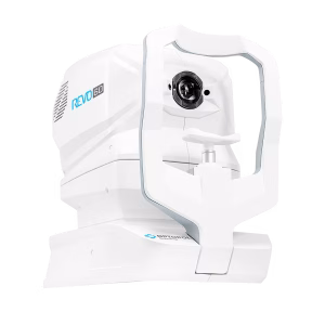
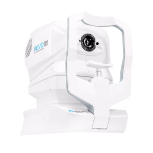
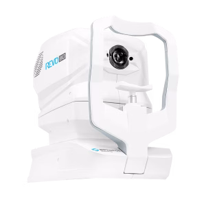
REVO HR


Ultra-High Resolution icon
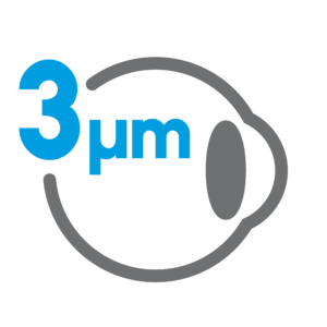
Ultra-High Resolution (3 µm)

The system provides 3 µm resolution imaging, enabling detailed visualization of fine ocular structures and improved detection of subtle pathological changes.


Auto Functions iconAuto Functions

Simplifying operation with the push of a button to auto-postion, auto-align, auto-focus, and auto-capture.


Structure & Function iconStructure&Function (S+F)

Comprehensive glaucoma solution that combines REVO OCT and PTS Visual Field results. S+F takes the diagnostic approach of the Hood report.Our S+F report allows clinicians to understand the relationship between structural glaucoma damage and the functional impact on the patient’s field of vision. This provides a quick and comprehensive single-page report for glaucoma management.


AccuTrack iconAccuTrack™

Our hardware-based eye tracker, compensates for blinks, loss of fixation and involuntary eye movements during scans reducing artifacts.


Glaucoma Tool Kit iconGlaucoma Tool Kit

Comprehensive glaucoma analytical tools for quantification of the Nerve Fiber Layer and Ganglion Cell Layer. The Disc Damage Likelihood Scale enables clinicians to precisely diagnose and monitor glaucoma for Optic Nerve Head analysis.


Full range iconFull range

With scans presenting New Extended Depth™ software, based on our Full Range technology, this new imaging mode provides scans of increased depth for reliable and convenient observation of challenging cases. The Full Range mode is perfect for diagnosing even highly myopic patients.


Progression Analysis iconProgression Analysis

Quickly view a chronological set of exams for analysis of changes in morphology, quantified progression maps, and progression trends.


Custom Scan Protocols iconCustom Scan Protocols

Save time and never miss a scan. Combine any scan type into a pre-set group. Choose a group of scans and set the order, the REVO will do the rest.


AI DeNoise iconAI DeNoise

An advanced artificial intelligence (AI) algorithm removes noise from the tomogram for the highest image quality.


Technical Specifications

FUNDUS CAMERA
Type Non-mydriatic fundus camera
Photography type Color
Angle of view 45° ± 5%
Min. pupil size for fundus 3.3 mm
Camera 12.3 Megapixel
Photography Fundus (Retina, Central, Disc, Peripheral with Manual fixation), Anterior
Flash adjustment, Gain, Exposure Auto, Manual
Intensity levels High, Normal, Low
OPTICAL COHERENCE TOMOGRAPHY
Technology Spectral Domain OCT
Light source SLED 870 nm, 93 nm width
Bandwidth 93 nm half bandwidth
Scanning speed 130 000 A-scan/sec
Axial resolution ~1.3 μm digital, 3 μm in tissue
Transverse resolution 12 μm, typically 18 μm
Overall scan depth 2.6 mm / 5.4 mm in Full Range mode
Min. pupil size for OCT 1.7 mm
Focus adjustment range -25 D to +25 D
Scan range Posterior 3–15 mm, Angio 3–15 mm, Anterior 3–18 mm
Scan types 3D, Angio², Full Range Radial, Full Range B-scan, Radial (HD), B-scan (HD), Raster (HD), Raster 21 (HD), Cross (HD), TOPO², Biometry AL²
Fundus alignment IR, pSLO (Live Fundus Reconstruction)
Alignment method Fully automatic, Automatic, Manual
Fundus tracking ACCUtrack – active real time, iTracking
Retina analysis Retina thickness, Inner Retinal thickness, Outer Retinal thickness, RNFL+GCL+IPL thickness, GCL+IPL thickness, RNFL thickness, RPE deformation, MZ/EZ-RPE thickness
Angiography OCT² Vitreous, Retina, Choroid, Superficial Plexus, RPCP, Deep Plexus, Outer Retina, Choriocapilaries, Depth Coded, SVC, DVC, ICP, DCP, Custom, Enface Quantification: FAZ, VFA, NFA, Vessel Area Density, Skeleton Area Density, Thickness map
Glaucoma analysis RNFL, ONH morphology, DDLS, OU and Hemisphere asymmetry, Ganglion analysis as RNFL+GCL+IP and GCL+IPL, Structure + Function¹
Angiography mosaic Acquisition: Auto, Manual; Mosaic modes: 10×10, 10×6, 12×5, 7×7, Manual up to 12 images
Biometry OCT² AL, CCT, ACD, LT, P, WTW
IOL Calculator³ Hoffer Q, Holladay I, Haigis, Theoretical T, Regression II
Corneal Topography Map² Axial [Anterior, Posterior], Refractive Power [Kerato, Anterior, Posterior, Total], Net Map, Axial True Net, Equivalent Keratometer, Elevation [Anterior, Posterior], Height, KPI (Keratoconus Prediction Index)
Anterior (no lens/adapter required) Anterior Chamber Radial, Anterior Chamber B-scan, Pachymetry, Epithelium map, Stroma map, Angle Assessment, AIOP, AOD 500/750, TISA 500/750, Angle to Angle view
Connectivity DICOM Storage SCU, DICOM MWL SCU, CMDL, Networking
Fixation target OLED display (the target shape and position can be changed), External fixation arm
Dimensions (L×W×H) / Weight 479 × 367 × 493 mm / 30 kg
Power supply / Consumption 100–240 V, 50/60 Hz / 90–110 VA
UWF LENS ⁴
Scan angle ~105° ⁵
Working distance 15 mm
Scan types 3D, Radial, Line, Angio², Full Range Radial, Full Range Line
Fundus tracking iTracking
Overall scan depth 2.6 mm / 5.4 mm in Full Range mode
¹ Via connection with PTS software version 3.4 or higher ² An optional software module ³ The Biometry module and a separate license for the IOL Calculator are required ⁴ Ultra-Wide Field imaging is available with the optional UWF lens ⁵ Measured from the center of the eye

You may also like…