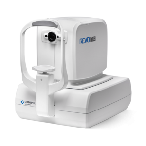
Description
New Model 80 000 A-scan/seconds
The new REVO model with 80 000 A-scan/sec scanning speed reduce scan time and brings benefits for both clinicians and patients by reducing errors often caused by involuntary eye movements. Higher sensitivity spectrometer allows to visualize finer details.
RETINA
Single 3D Retina examination is enough to perform both Retina and Glaucoma analysis based on retinal scans. Software automatically recognizes 8 retina layers. Thusallowingamoreprecisediagnosisandmapping of any changes in the patient’s retina condition.

The SOCT Copernicus REVO 60 000 and 80 000 A-scan/seconds is available now with the Angiography module.
OCT Angiography
This module allows visualization of the retinal microvasculature. Angiography SOCT is a non-invasive, dye-free technique providing 3D image of retinal blood circulation.
| Standard Single View | Detailed Single View |
 |  |
| Both View | |
 |
| Comparison View | Progression View |
 |  |
QUANTYFICATION TOOLS: FAZ, VFA
FAZ – Foveal Avascular Zone measurements allow to quantify and monitor changes in Superficial and Deep vascular layer. FAZ tool is also available for narrow and wide scans.
 |  |
| Area [mm2]: 0,48 |
VFA – Vascular Flow Area allows to examine the pathologically affected area and precisely measure the area covered by vascu-larization. User can easily measure area on predefined or own selected vascular layer.
 |  |
| Area [mm2]: 13,45 Flow area [mm2]: 2,85 |
ANGIO MOSAIC
The Angiography mosaic delivers high-detailed images over large field of the retina. Advanced tab provides: view of any vascular layers, enface view of vascular layers, depth coded and thickness map.
Mosaic modes: 10×6 mm


Manual (up to 12 images)

GLAUCOMA
Comprehensive glaucoma analytical tools for quantification of the Nerve Fiber Layer, Ganglion layer and Optic Head with DDLS allow for the precise diagnosis and monitoring of glaucoma over time.
With the golden standard 14 optic nerve parameters and a new Rim to Disc and Rim Absence the description of ONH condition is quick and precise.
Advanced view which provides combined information from Retina and Disc scan to integrate details of the Ganglion cells, RNFL, ONH in a wide field perspective for comprehensive analysis.
| Advance Retina & ONH | ONH Single |
 |  |
Asymmetry Analysis of Ganglion layers between hemi-spheres and between eyes allows easier identification and detection of glaucoma in early stages and in non-typical patients.
| Ganglion Both | Ganglion Progression |
 |  |
Implemented the DDLS – Disc Damage Likelihood Scale which use 3 separate classification for small, average and large discs. It supports the practitioners in a quick and precise evaluation of the patient’s glaucomatous disc damages.
| ONH Both | ONH Progression |
 |  |
COMPLET YOUR GLAUCOMA REPORT
To eliminate common problem with the understanding of the patient’s IOP pachymetry module provides IOP Correction value. With the implemented Adjusted IOP formula you can quickly and precisely understand the measured IOP value.
As the Pachymetry and Anterior Chamber Angle Verification require no additional attachments, the predefined Glaucoma protocol, which consists of Retina, Disc and Anterior scans, can be done automatically to reduce patient chair time.
| Closing angle | Anterior single view |
 |  |
| * Images courtesy of Prof. Edward Wylęgała MD, PhD | |
ANTERIOR
For standard examination no additional lens is required. Additional adapter provided with the device allows to make wide scans of anterior segment.
| Cornea single | Cornea both |
 |  |
| Cornea Comparison | Cornea Progression |
 |  |
BIOMETRY OCT is optional software module to purchase for REVO model with 27 000, 60 000 nad 80 000 A-scan/sec scanning speed
B-OCT™ innovative method of using the posterior OCT device to measure ocular structure along eye axis .
OCT Biometry provides complete set of Biometry parameters: Axial Length AL, Central Cornea Thickness CCT, Anterior Chamber Depth ACD, Lens Thickness LT.
Result review | Analysis window |
Visually verify your measurement
All measurement calipers are shown on the all boundaries OCT image provided by REVO. It allows to can visually verify and correct what structure of the eye has been measured.
TOPOGRAPHY OCT is optional software module to purchase
T-OCT™ is a pioneering way to provide detailed corneal Curvature maps by using posterior dedicated OCT. Ante-rior, Posterior surface and Corneal Thickness allow to provide the True Net Curvature information. With Net power, the precise understading of the patient’s corneal condition comes easily and is free of errors associated with modelling of posterior surface of the cornea. SOCT T-OCT module provides Axial maps, Tangential maps, Total Power map, Height maps, Epithelium and Corneal thickness maps.
Corneal topography module clearly shows the changes in the cornea on the difference map view. Customize your
Topography module provides:
- Full featured Corneal mapping of Anterior, Posterior and Real
- Precise Astigmatism Display Option (SimK: Anterior, Posterior, Real, Meridian and Emi-Meridian ø 3, 5, 7 mm zones
KERATOCONUS SCREENING
Easly detect and classified keratoconus with Keratoconus classifier. Classification based on KPI, SAI, DSI, OSI and CSI. In the early stages of kera-toconus the results can be complemented by Epithelium and Pachymatery maps.
| Normal | Astigmatysm | Keratoconus |
 |  |  |
COMPARE THE EXAMS
Comprehensive software features a range of selectable views: Single, Both. See details on standard Singe view and easly see corneal asymmetry on the Both view.
The follow-up feature in to the T-OCT™ module, allows fully compare the changes in the corneal topography over time for:
- LASIK undergone patients
- Keratoconus patients
- The contact lens wearers
| Single | Both |
 |  |
| Comparison | Progression |
 |  |
FOLLOW UP
High density of standard 3D scan allows to precisely track the disease progression. Operator can analyze changes is morphology, quantified progression maps and evaluate the progression trends.
| Progression Morphology | Progression Quantification |
 |  |
COMPARISON
Comparison view allows in easy and convince way to review results from two visits. The view is available for most of the scan programs.

NETWORKING
A proficient networking solution increases productivity and an enhanced patient experience. It allows you to view and manage multiple examinations from review stations in your practice. Effortlessly helping to facilitate patient education by allowing you to interactively show examination results to patients. Every practice will have different requirements which we can provide by tailoring a bespoke service. There is no additional charge for the server module.
DICOM
Store, exchange, and transmit results through DICOM gateway to the hospital network.
ENHANCE TOMOGRAMS – 2 µm Ultra High (Digital) Resolution

CHOROID VISUALIZATION

CLINICAL IMAGES


CORNEA SCAN

Wide cornea scan, Descemet’s membrane detachment (DMD) and iridocorneal adhesions

* Images courtesy of Prof. Edward Wylęgała MD, PhD
BACK OF THE CRYSTALLINE LENS

WIDE SCAN WITH LENS DEFECT

CONTACT LENS ON THE CORNEA

CONTACT LENS ON THE CORNEA AND THE SCLERA

ANTERIOR WIDE

Angle to Angle scan, narrow angles

*Image courtesy of Bartosz L. Sikorski MD, PhD
WIDE RETINA SCAN

PERIPHERAL SCAN