

Description
Imaging at the cellular level
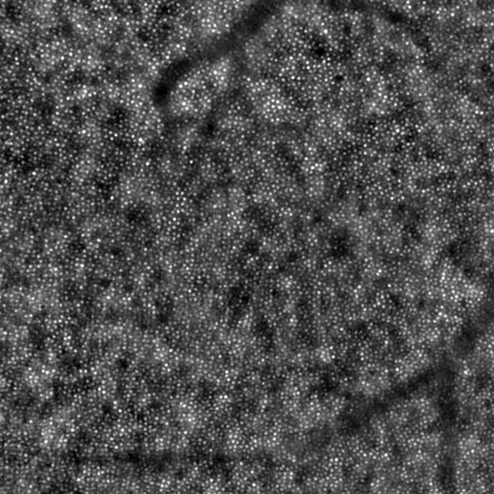
When using the rtx1 Adaptive Optics Retinal Camera, you examine the retina at a scale where individual cells are visible. Its ultrahigh-resolution images reveal parafoveal cone photoreceptors as well as other microscopic retinal structures that cannot be seen with conventional techniques.
Microvascular imaging
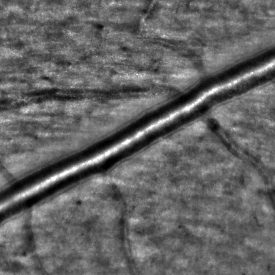
The rtx1 enables to visualize the microscopic walls of retinal arterioles non-invasively. Focal narrowing, perivascular sheathing, micro-hemorrhages and micro-aneurisms are also visible without using contrast agents.
Track microscopic progression/regression
By design, the rtx1 delivers images that are free from motion distortion. Building on this advantage, its software enables capturing the same retinal region through different visits, and automatically aligns follow-up images. This allows tracking minute changes in a group of cells, a vessel section, or a lesion over time.
Image credit: Quinze-Vingts National Eye Hospital, Paris, France


