Description
Spectral-Domain OCT
Fully automated capture with the push of the START button.
Moving OCT innovation forward. OPTOPOL pioneered the first Spectral Domain OCT technology 18 years ago. Our background allows us to provide an unparalleled experience to both patients and eye care professionals. Get the best of both worlds with ultra-high quality scans in an easy-to-use interface.
- Provides both Posterior and Anterior scan capabilities.
- Combine one-touch exams with preset scan combinations for streamlined workflows.
- Progression analysis available for detailed analysis over time.
- Add-on software modules to increase the efficiency of your OCT device.
Non-Myd Color Fundus Camera
Capture high-resolution 45° true color retinal imaging.
The REVO FC OCT series adds a 12.3 MP Fundus Camera, capable of capturing ultra-high quality color images. The REVO FC series Fundus Camera is fully automated and easy to use.
- Auto-alignment, auto-focus, auto-flash, auto-capture with the push of a button.
- Capture high resolution fundus images with a pupil size as small as 3.3 mm.
- Toolkit of various fundus image processing tools.
- Deliver detailed photos of one or both eyes as well as a chronological comparison.
- Save time by attaching a single fundus photo to several OCT scans.
Take your OCT to the next level
REVO FC 130
AccuTrack™
Our hardware-based eye tracker, compensates for blinks, loss of fixation and involuntary eye movements during scans reducing artifacts.
Auto Functions
Simplifying operation with the push of a button to auto-postion, auto-align, auto-focus, and auto-capture.
AI DeNoise
An advanced artificial intelligence (AI) algorithm removes noise from the tomogram for the highest image quality.
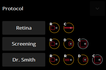
Custom Scan Protocols
Save time and never miss a scan. Create a custom preset group of scans and let the REVO capture all scans in order.
Motion Correction
The software-based motion correction (MC) compensates for involuntary eye movements and blinks by capturing two scans and generating a motion corrected scan when necessary.
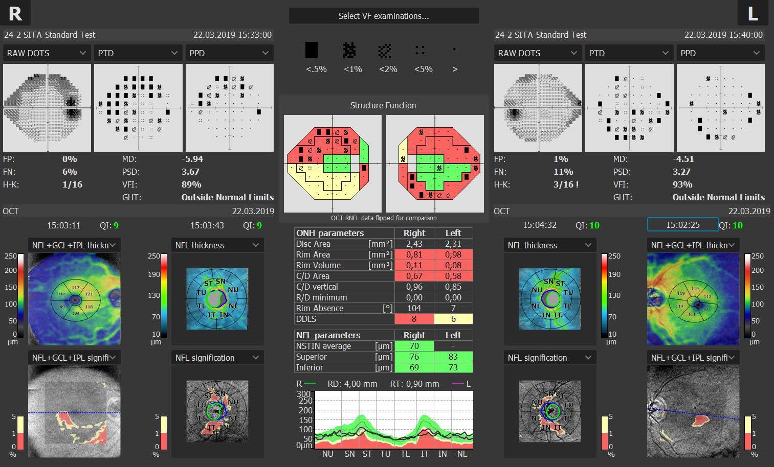
Structure + Function (S+F)
Comprehensive glaucoma solution that combines REVO OCT and PTS Visual Field results. S+F takes the diagnostic approach of the Hood report.
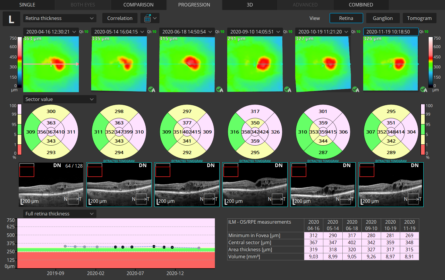
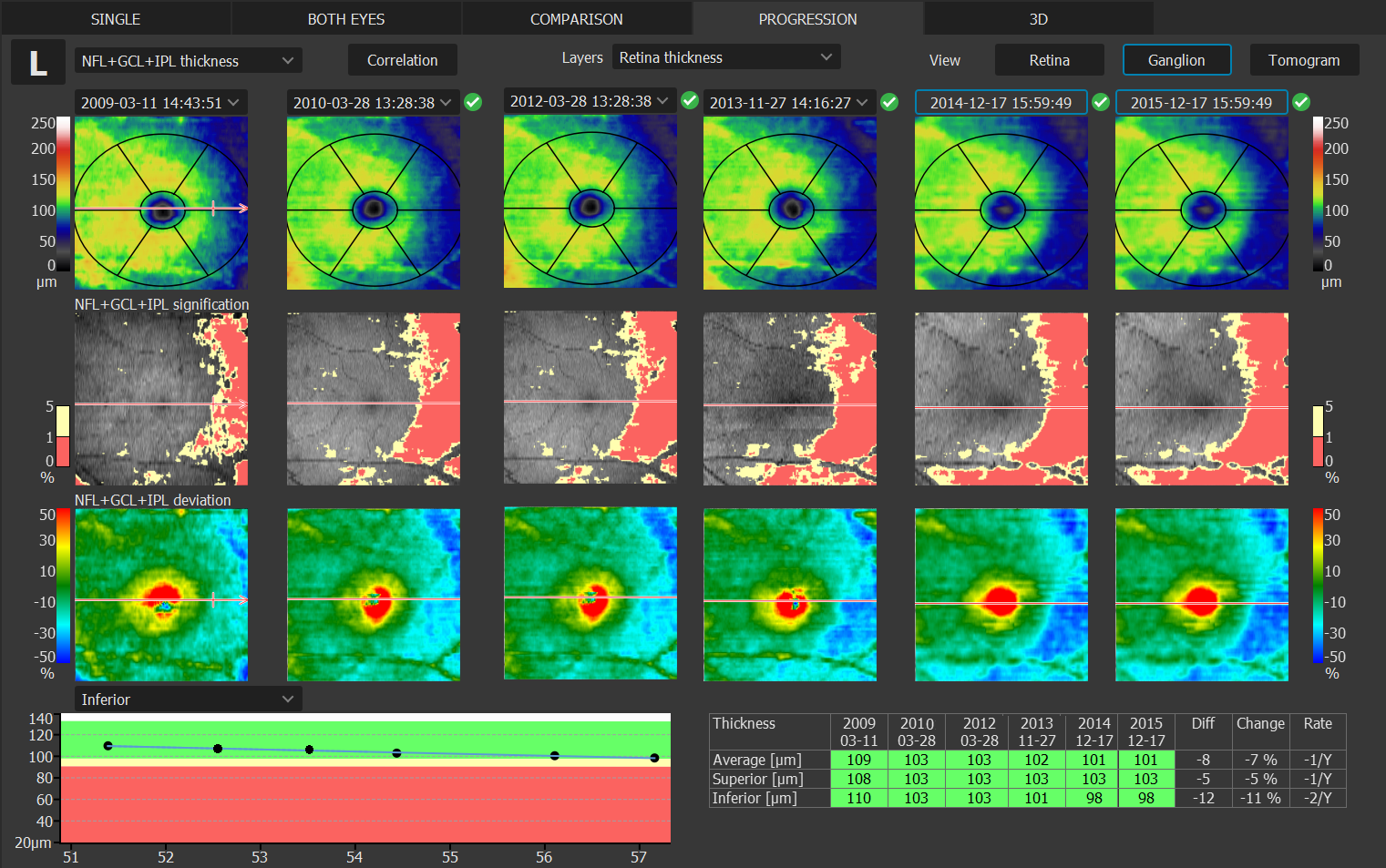
Progression Analysis
Gather baselines and follow-ups to monitor and manage disease progression in posterior and anterior scans.
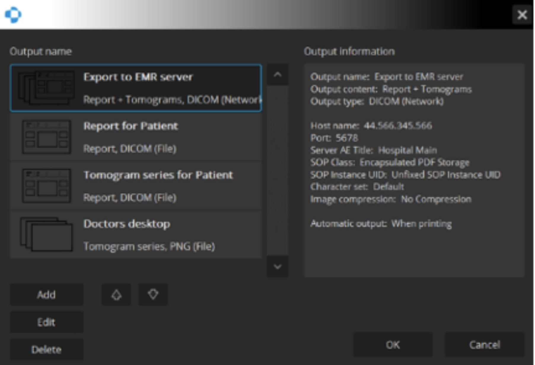
Connectivity
A proficient networking solution with DICOM and EMR capabilities. Quickly and easily export to a desired location.
FOLLOW DISEASE PROGRESSION
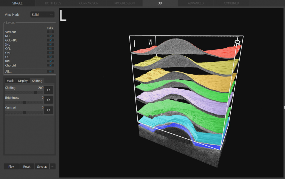
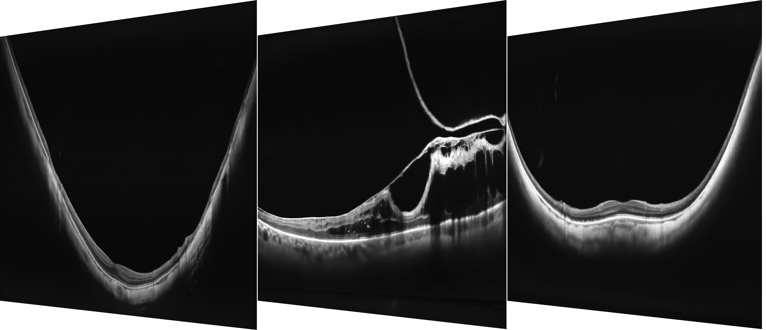
Full Range
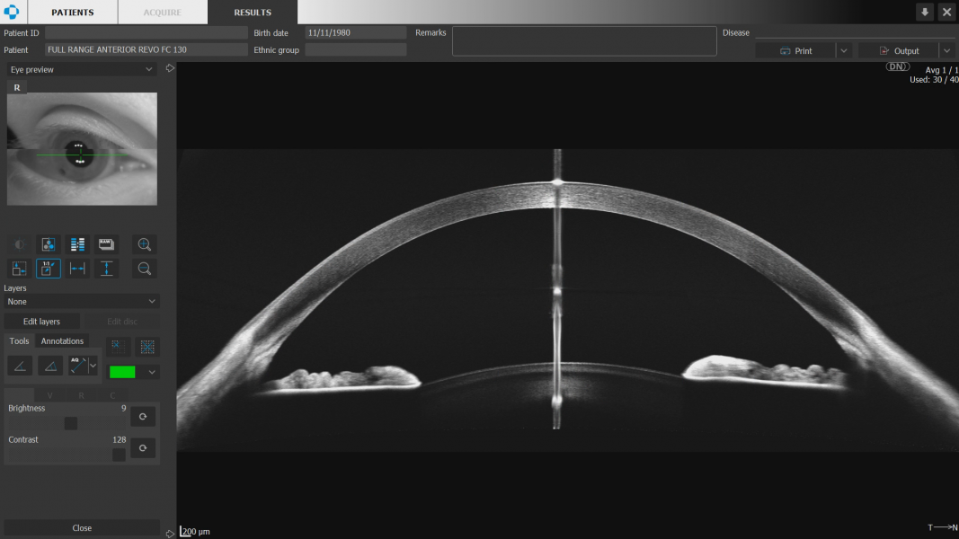
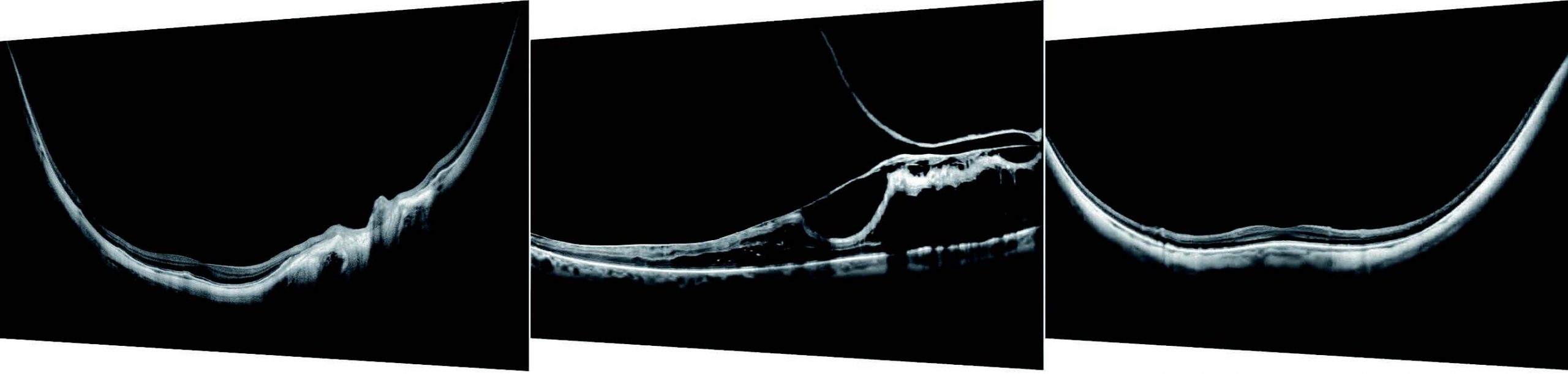
With scans presenting New Extended DepthTM software, based on our Full Range technology, provides scans of increased depth for reliable and convenient observation of challenging cases. With scans presenting extended depth, this new imaging mode is perfect for diagnosing even highly myopic patients.
STANDARDIZE SCANS PERFORMED
Connectivity
REVO offers an excellent networking solution that allows doctors to view and manage multiple examinations from review stations in your practice. The system comes with the latest version of Windows PRO and can easily export in PDF, JPEG and DICOM formats to your network or EHR.
FOLLOW DISEASE PROGRESSION
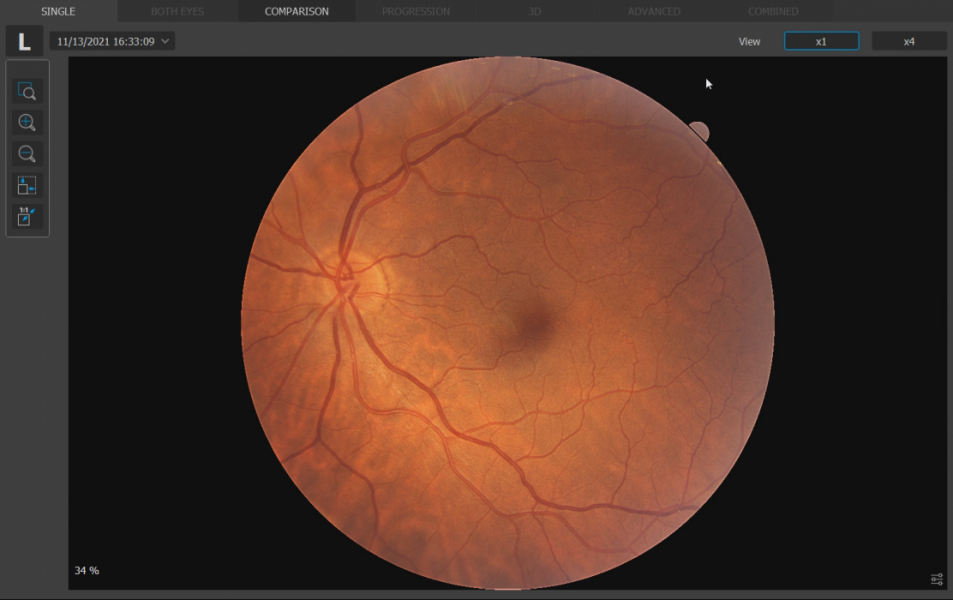
Progression Analysis
Quickly view a chronological set of exams for analysis of changes in morphology, quantified progression maps, and progression trends.
STANDARDIZE SCANS PERFORMED
Custom Scan Protocols
Save time and never miss a scan. Combine any scan type into a pre-set group. Choose a group of scans and set the order, the REVO will do the rest.
FOLLOW DISEASE PROGRESSION
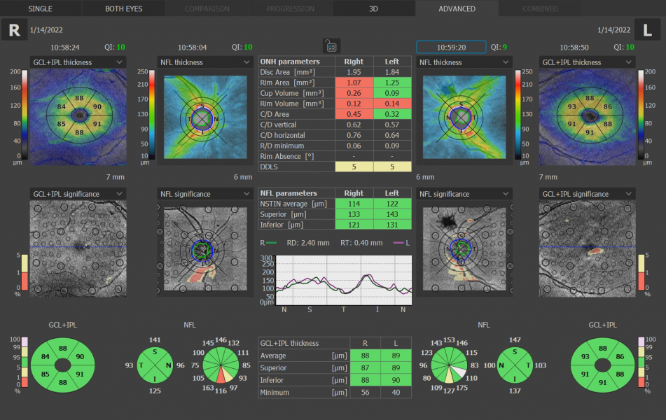
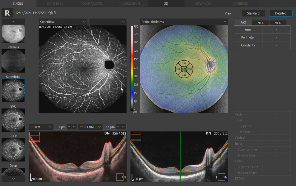
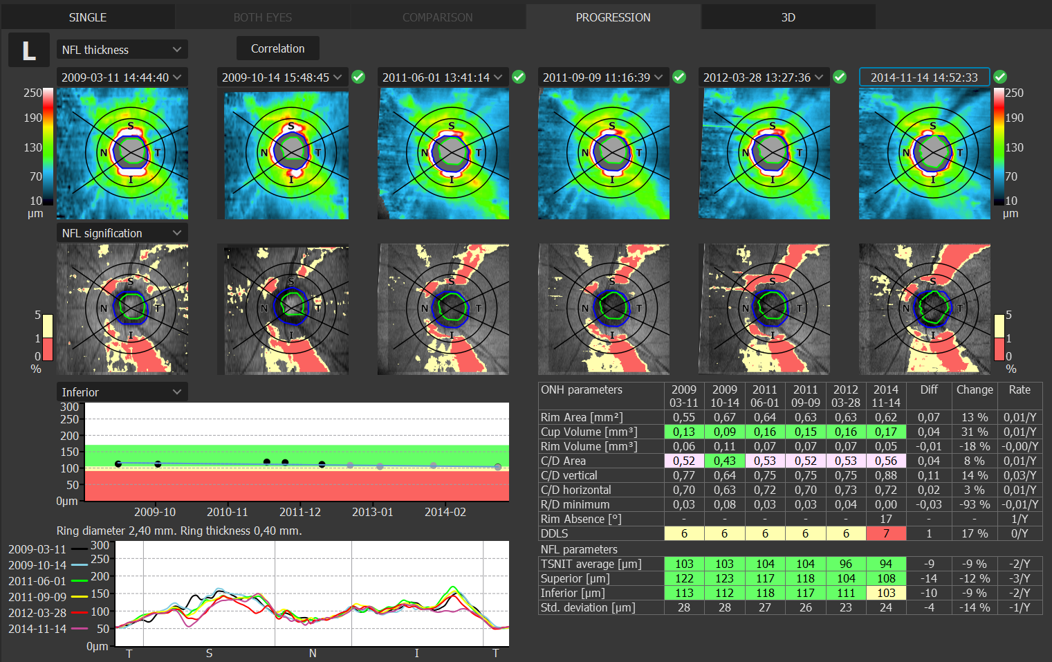
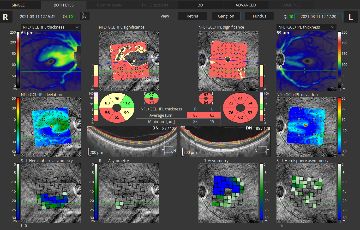
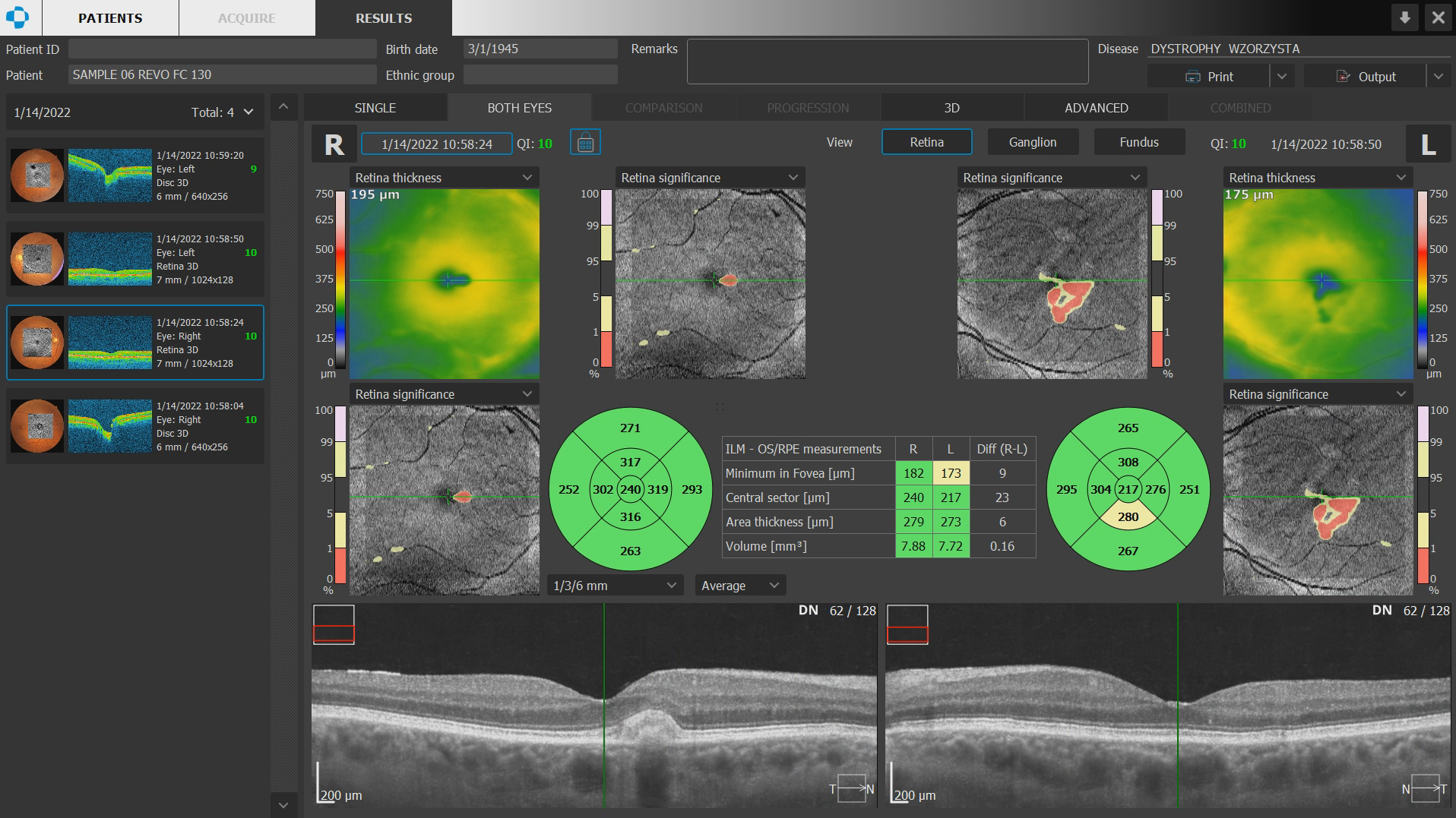
Glaucoma Tool Kit
Comprehensive glaucoma analytical tools for quantification of the Nerve Fiber Layer and Ganglion Cell Layer. The Disc Damage Likelihood Scale enables clinicians to precisely diagnose and monitor glaucoma for Optic Nerve Head analysis.
COMPREHENSIVE HOOD REPORT
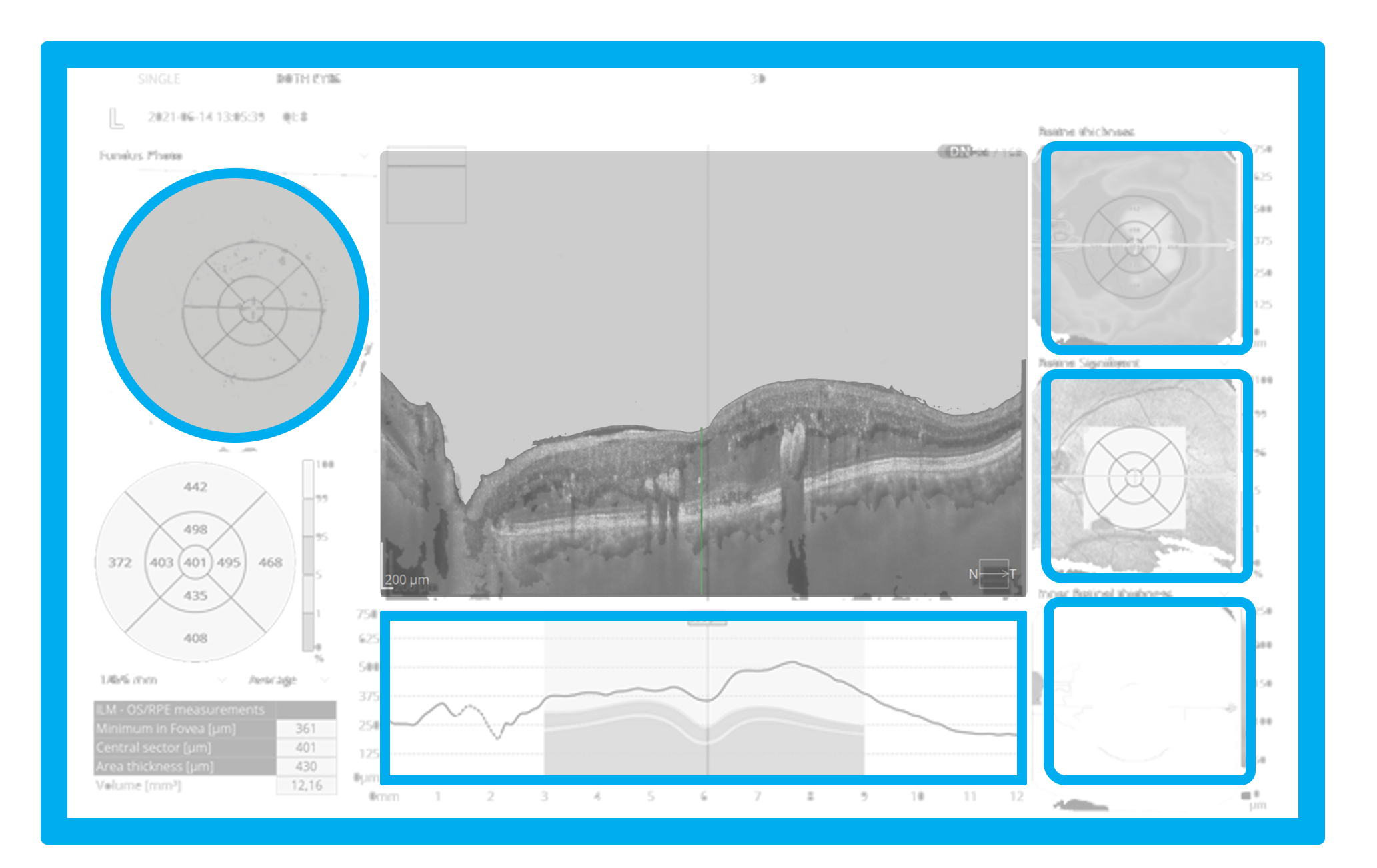
Structure + Function
Our S+F report allows clinicians to understand the relationship between structural glaucoma damage and the functional impact on the patient’s field of vision. This provides a quick and comprehensive single-page report for glaucoma management.
Perform high-resolution posterior and anterior segment scans
Additional Scan Programs
Retina
- 3D Volume Scan
- Radial Line Scan
- Raster Line Scan
- Retina Cross Scan
- High Resolution Line Scan
Disc
- 3D Volume Scan
- Radial Line Scan
- High Resolution Line Scan

Wide Field
- 3D Volume Scan
- Raster Line Scan
- Full Range Line Scan
Anterior
- 3D Volume Scan
- Radial Line Scan
- Raster Line Scan
- Anterior Cross Scan
- Anterior Line Scan
- Anterior Chamber Full-Range Radial Scan
- Anterior Chamber Full-Range Line Scan