Description
Spectral-Domain OCT
Fully automated capture with the push of the START button.
Moving OCT innovation forward. OPTOPOL pioneered the first Spectral Domain OCT technology 25 years ago. Our background allows us to provide an unparalleled experience to both patients and eye care professionals. Get the best of both worlds with ultra-high quality scans in an easy-to-use interface.
- Provides both Posterior and Anterior scan capabilities.
- Combine one-touch exams with preset scan combinations for streamlined workflows.
- Progression analysis is available for detailed analysis over time.
- Add-on software modules to increase the efficiency of your OCT device.
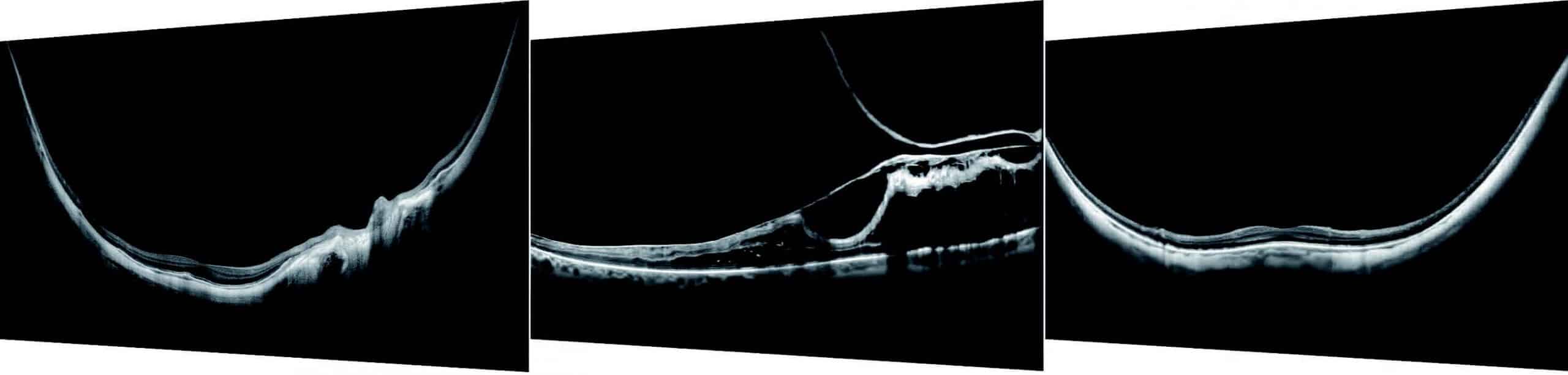
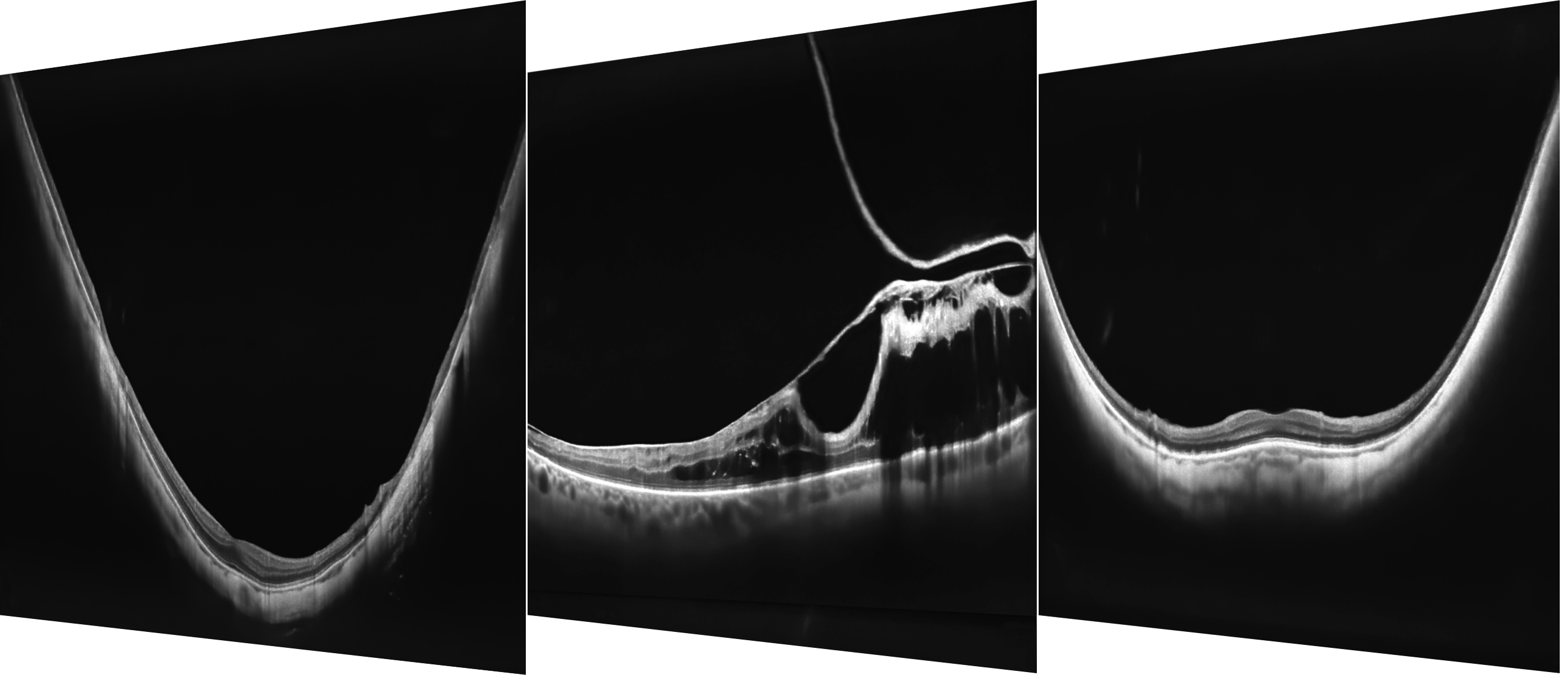
Full Range
With scans presenting New Extended DepthTM software, based on our Full Range technology, provides scans of increased depth for reliable and convenient observation of challenging cases. With scans presenting extended depth, this new imaging mode is perfect for diagnosing even highly myopic patients.
STANDARDIZE SCANS PERFORMED
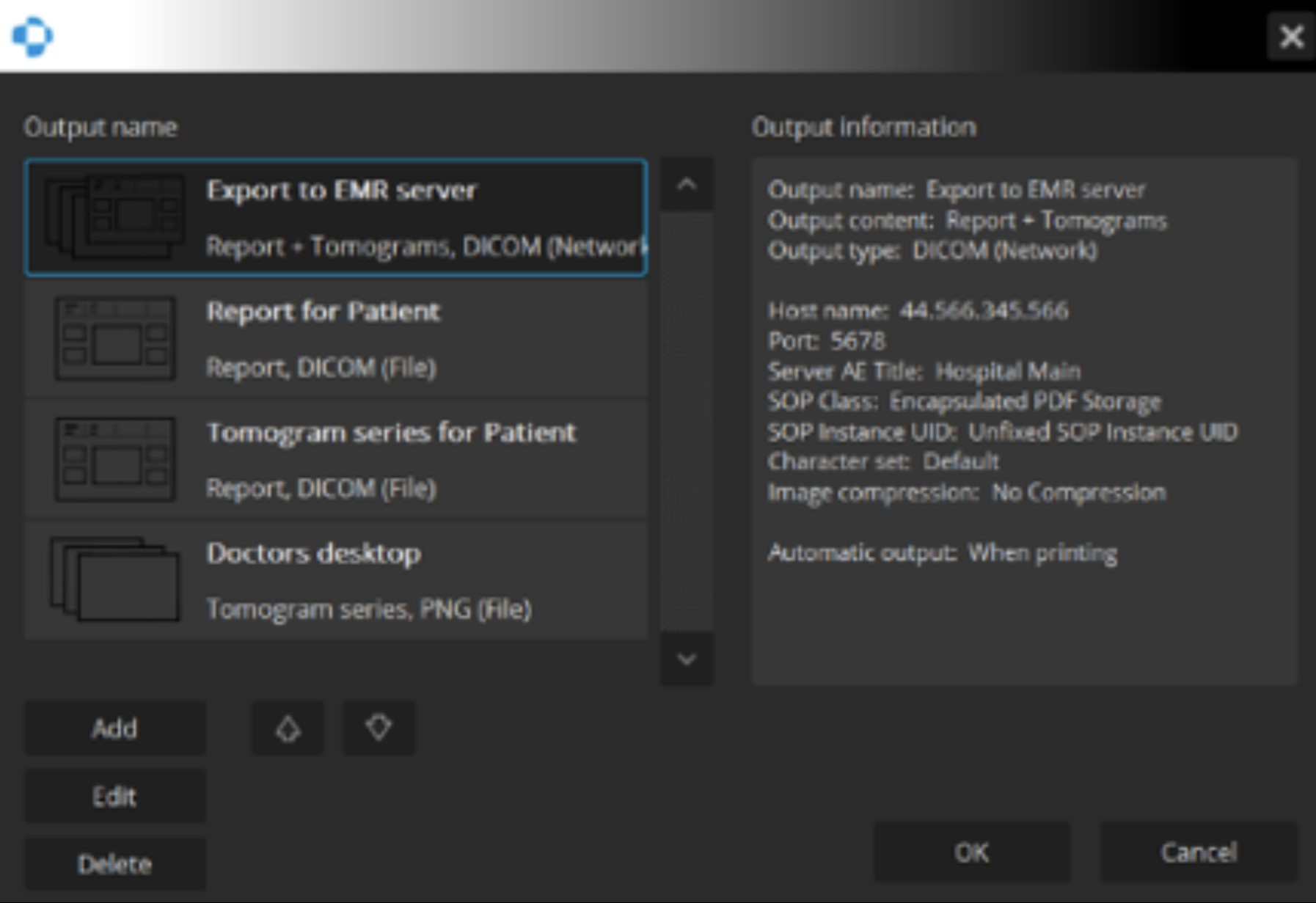
Connectivity
REVO offers an excellent networking solution that allows doctors to view and manage multiple examinations from review stations in your practice. The system comes with the latest version of Windows PRO and can easily export in PDF, JPEG and DICOM formats to your network or EHR.

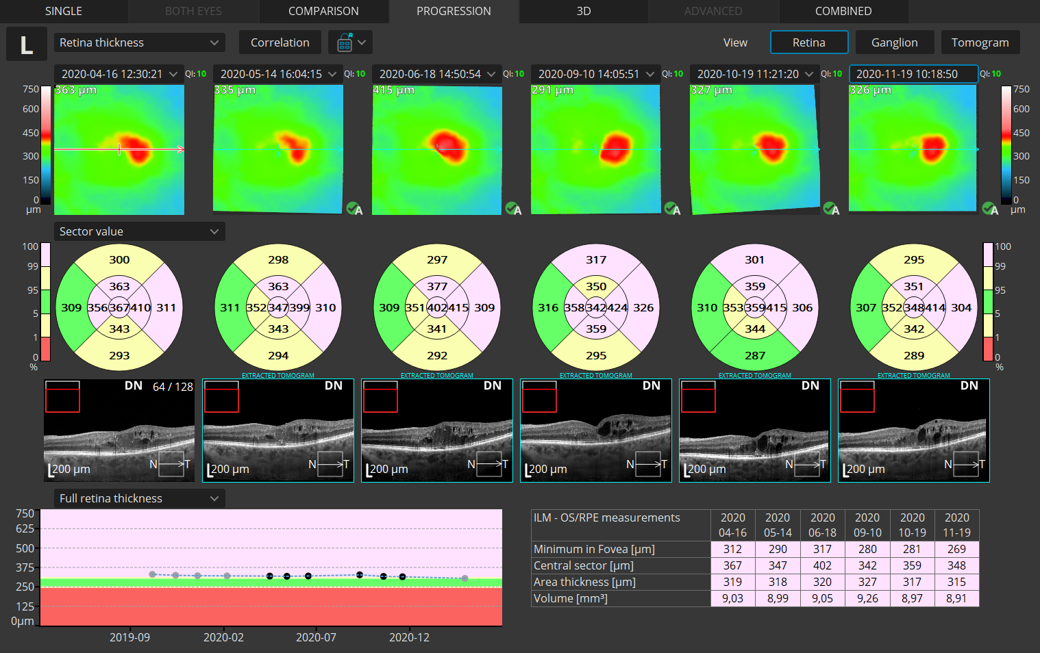
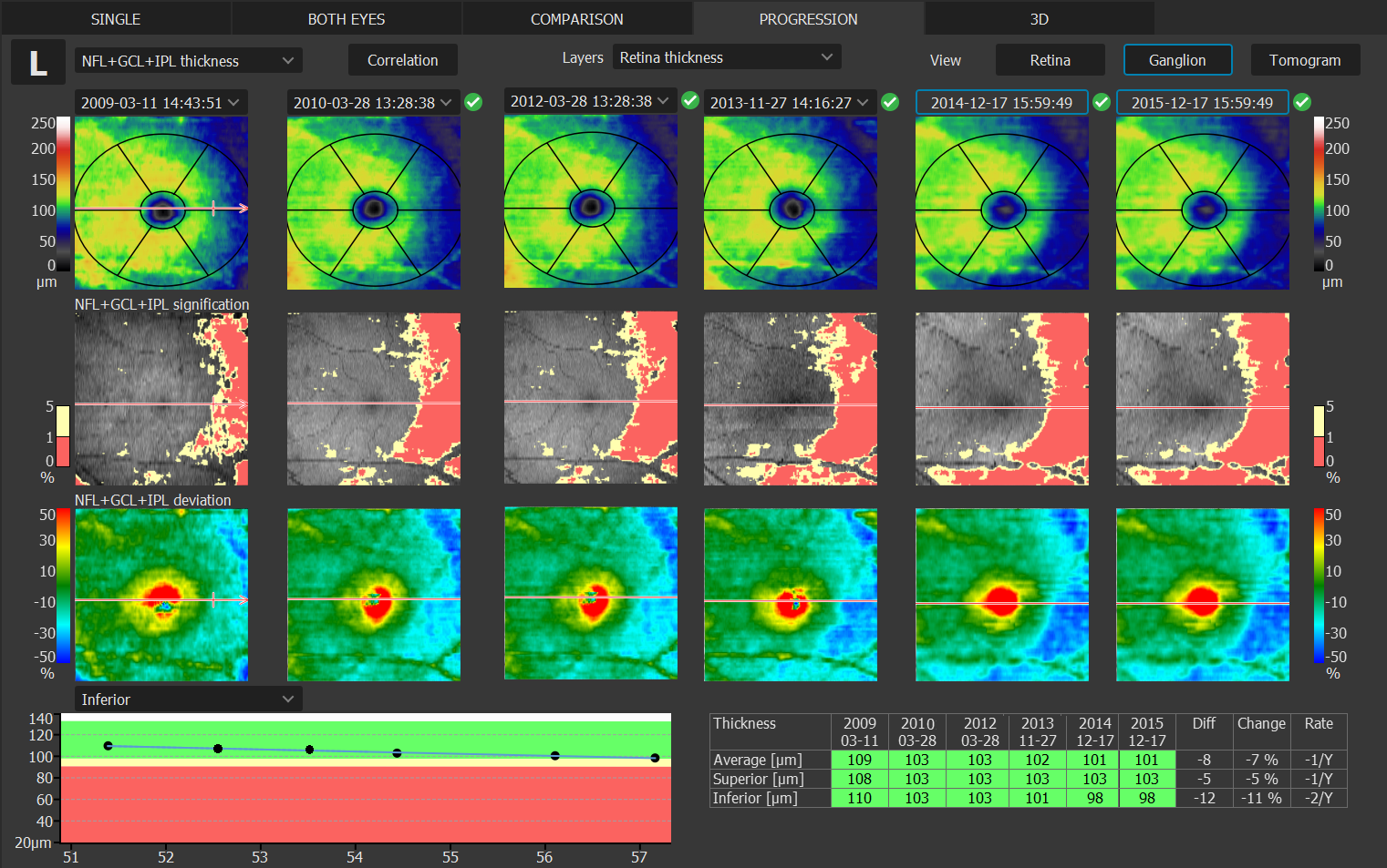
FOLLOW DISEASE PROGRESSION
Progression Analysis
Quickly view a chronological set of exams for analysis of changes in morphology, quantified progression maps, and progression trends.
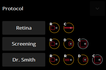
STANDARDIZE SCANS PERFORMED
Custom Scan Protocols
Save time and never miss a scan. Combine any scan type into a pre-set group. Choose a group of scans and set the order, the REVO will do the rest.
FOLLOW DISEASE PROGRESSION
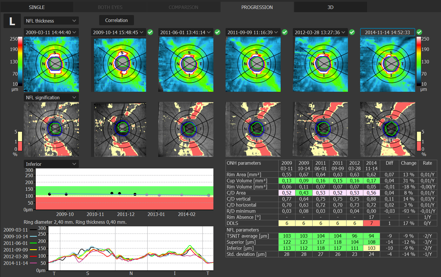
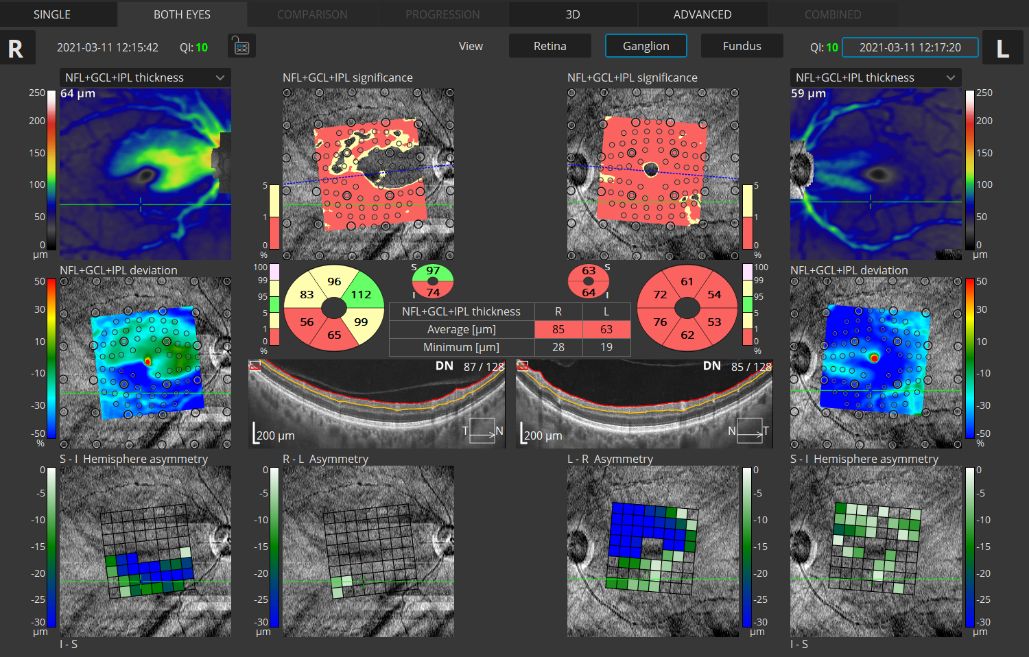
Glaucoma Tool Kit
Comprehensive glaucoma analytical tools for quantification of the Nerve Fiber Layer and Ganglion Cell Layer. The Disc Damage Likelihood Scale enables clinicians to precisely diagnose and monitor glaucoma for Optic Nerve Head analysis.
COMPREHENSIVE HOOD REPORT
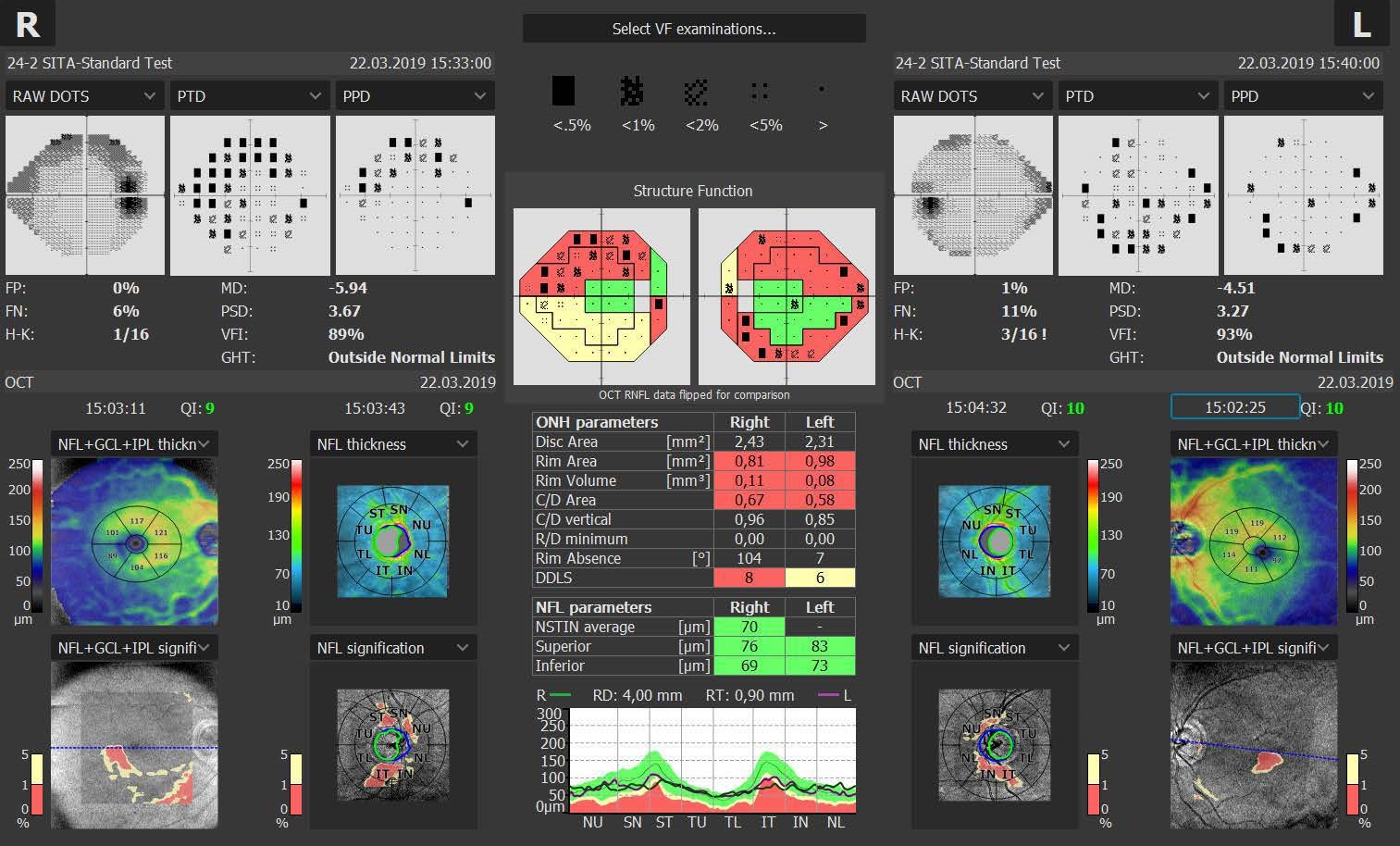
Structure + Function
Our S+F report allows clinicians to understand the relationship between structural glaucoma damage and the functional impact on the patient’s field of vision. This provides a quick and comprehensive single-page report for glaucoma management.
Additional Scan Programs
Perform high-resolution posterior and anterior segment scans
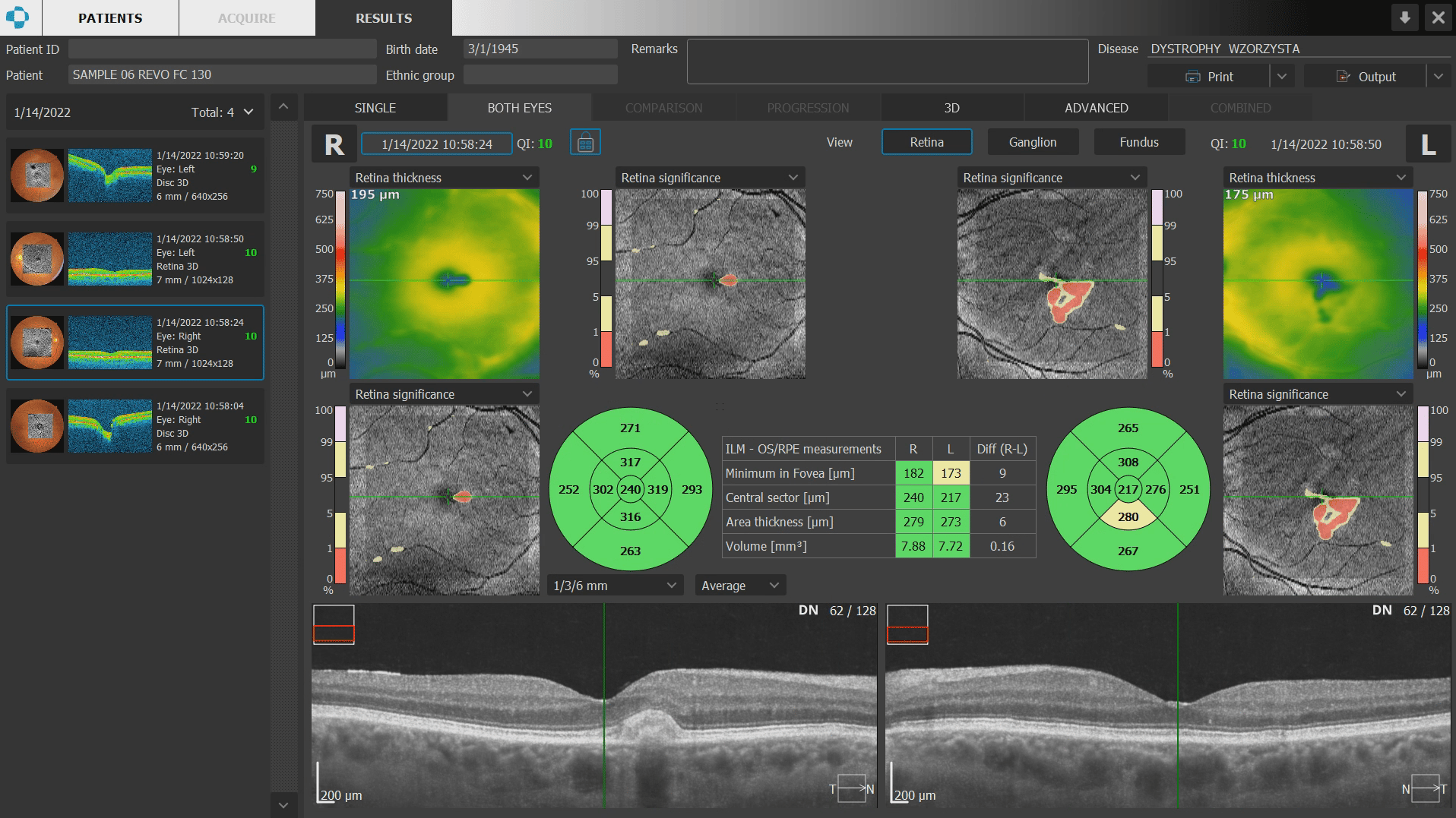
Retina
- 3D Volume Scan
- Radial Line Scan
- Raster Line Scan
- Retina Cross Scan
- High Resolution Line Scan
Disc
- 3D Volume Scan
- Radial Line Scan
- High Resolution Line Scan

Wide Field
- 3D Volume Scan
- Raster Line Scan
- Full Range Line Scan
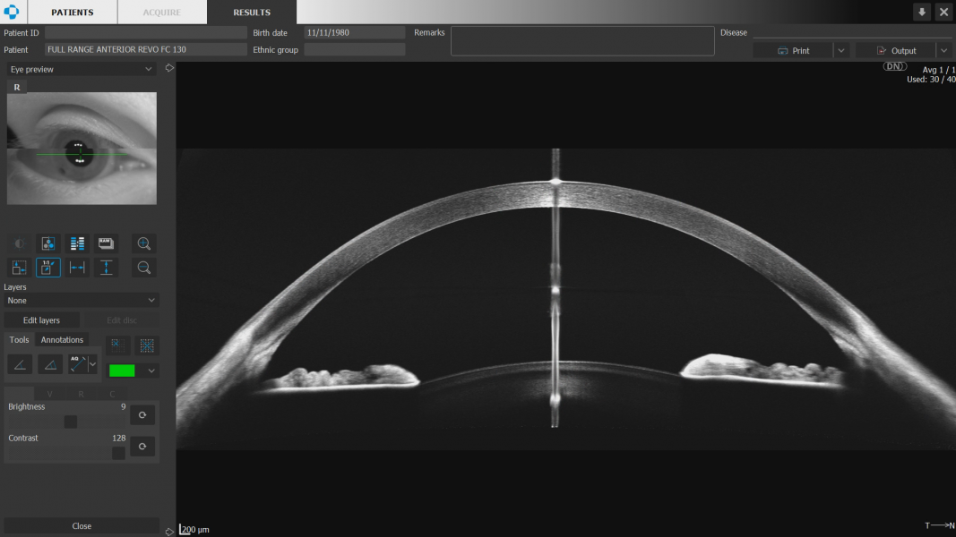
Anterior
- 3D Volume Scan
- Radial Line Scan
- Raster Line Scan
- Anterior Cross Scan
- Anterior Line Scan
- Anterior Chamber Full-Range Radial Scan
- Anterior Chamber Full-Range Line Scan
Technical Specifications
| Technology |
Spectral Domain OCT
|
| Scanning speed | 60 000 measurements per second |
| Light Source |
SLED Wavelength 850 nm
|
| Bandwidth |
50 nm half bandwidth
|
| Axial resolution | 2.8 μm digital, 5 μm in tissue |
| Transverse Resolution |
12 μm, typical 18 μm
|
| Overall scan depth |
2.8 mm / ~6 mm in Full Range mode
|
| Scan range |
Posterior 5 mm to 15 mm, Angio 3 mm to 6 mm, Anterior 3 mm to 18 mm
|
| Scan program | 3D, Angio¹, Full Range Radial, Full Range B-scan, Radial, B-scan, Raster, Cross, TOPO¹ , Biometry AL¹ |
| Fundus image |
Live Fundus Reconstruction
|
| Alignment method |
Fully automatic, Automatic, Manual
|
| Retina analysis |
Retina thickness, Inner Retinal thickness, Outer Retinal thickness, RNFL+GCL+IPL thickness, GCL+IPL thickness, RNFL thickness, RPE deformation, MZ/EZ-RPE thickness
|
| Angiography OCT¹ | Vitreous, Retina, Choroid, Superficial Plexus, RPCP, Deep Plexus, Outer Retina, Choriocapilaries, Depth Coded, SVC, DVC, ICP, DCP, Custom, Enface, FAZ, VFA, NFA, Quantification: Vessel Area Density, Skeleton Area Density, Thickness map |
| Angiography mosaic | Acquistion method: Auto, Manual Mosaic modes: 10×6 mm, Manual up to 12 images |
| Glaucoma analysis |
RNFL, ONH morphology, DDLS, OU and Hemisphere asymmetry, Ganglion analysis as RNFL+GCL+IP and GCL+IPL,
Structure + Function² |
| Anterior |
Pachymetry, Epithelium map, Stroma map, AIOP, Angle Assessment, AOD 500/750, TISA 500/750
|
| Anterior (no lens/adapter required) |
Anterior Chamber Radial, Anterior Chamber B-scan, Angle to Angle view
|
| Biometry OCT | AL, CCT, ACD, LT, P, WTW |
| IOL Calculator ³ | IOL Formulas: Hoffer Q, Holladay I, Haigis, Theoretical T, Regression II |
| Corneal Topography Map¹ | Axial [Anterior, Posterior], Refractive Power [Kerato, Anterior, Posterior, Total], Net Map, Axial True Net, Equivalent Keratometer, Elevation [Anterior, Posterior], Height, KPI (Keratoconus Prediction Index) |
| Connectivity | DICOM Storage SCU, DICOM MWL SCU, CMDL, Networking |
| Min. pupil size | 2.4 mm |
| Focus adjustment range | -25D to +25D |
| Dimensions (LxWxH) / Weight | 479 mm x 367 mm × 493 mm / 29 kg |
| Fixation target | OLED display (The target shape and position can be changed), External fixation arm |
| Power Supply / Consumption | 100 V to 240 V, 50/60 Hz / 90 VA to 110 VA |
¹ An optional software module
² via connection with PTS software version 3.4 or higher
³ The Biometry module and a separate license for the IOL Calculator are required